You can do just about anything with touch-screen technology:
Play a game.
Respond to an email.
Dissect a body.
With a swipe of a finger, Shenandoah University students can now virtually dissect a body on an Anatomage life-size virtual dissection table, which uses data from real patient and cadaver scans to create a high-resolution 3D presentation.
The approximately $65,000 table arrived at Shenandoah’s Health Professions Building on the Winchester Medical Center campus over the summer, and a few Physician Assistant Studies (PA) program students were among the first to see it. A generous grant from the Shingleton Foundation provided the majority of funding for the table.
“Oh my gosh, this is so cool,” PA student Serena Schwechter Kalish ’17, of Silver Spring, Maryland, said as she encountered the table.
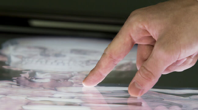
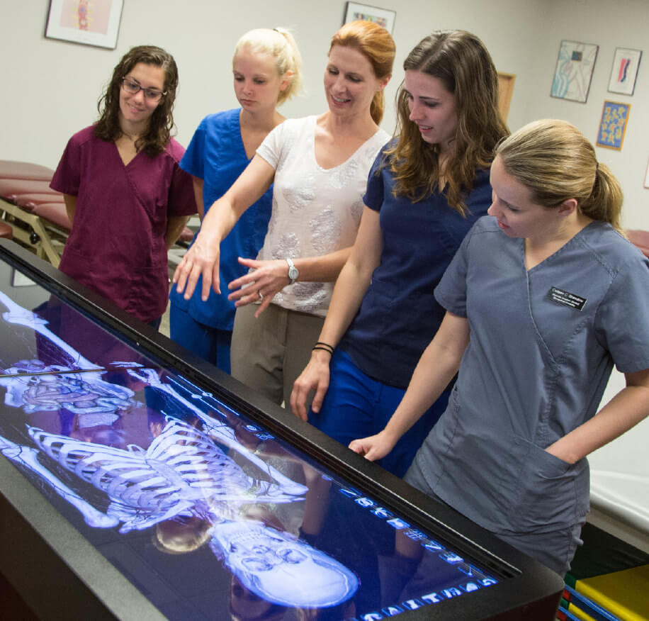
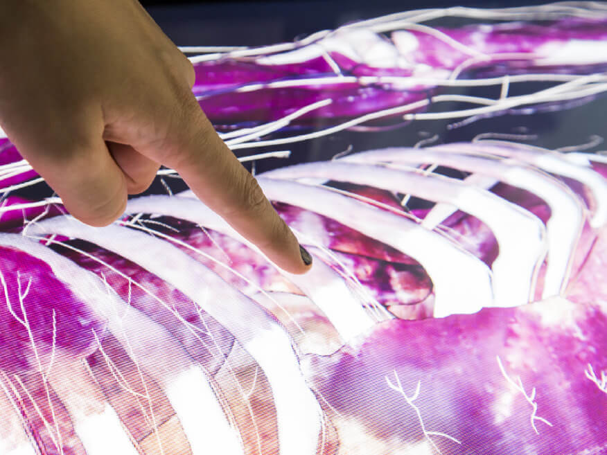

Assistant Professor of Physical Therapy Thomas Turner, Ph.D., who is an anatomy lab instructor, said that with the virtual device, you can turn a body from side to side, rotate the whole thing, and get a side view, look at a transection and more, all with the swipe of a finger. “With a simple touch of a couple of fingers, we can slide through different layers of the body,” he said.
“Virtual anatomy, which is what the Anatomage Table provides, is an excellent way for students to really learn human anatomy,” said Timothy Ford, Ph.D, dean of the School of Health Professions. “There will always be a place for the traditional anatomy lab, but this is the next best thing and is a technology that is increasingly being adopted by medical schools. This will lessen the pressure on the anatomy lab, allow students in health professions to practice their skills and knowledge in a relaxed setting at the Health Professions Building (which does not have an anatomy lab), and provide opportunities for interprofessional practice/exercises which are difficult to conduct in the traditional anatomy lab with all its necessary rules and regulations.
“It’s on wheels, so we can move it between classrooms, and transfer virtual dissections by VTC between campuses,” Dr. Ford continued. “It’s clean, odorless and looks like a human-sized iPad with all the functionality for complete human anatomy.”
The table offers access to life-size horizontal cross sections, and vertical sections that divide the body into left and right halves and from front to back. It also allows graduate health professions students to see where body structures are, Turner said. It’s a great adjunctive tool for an anatomy (cadaver) lab that allows users to do a cross-section on the spot, using a “scalpel” tool, something which cannot be done in an anatomy lab. The views available on the table are also helpful to those who will read CT and MRI scans, Turner said.
When a brain cross-section came into view on the table, Turner said, “That’s totally an MRI there.”
But it’s not the only tool for health professions students. Turner said the university also still maintains its cadaver labs, which provide students with great information, at its Health & Life Sciences Building in Winchester and Scholar Plaza at the Northern Virginia Campus in Loudoun County. The table is a great help — particularly as a teaching tool and when body systems need to be looked at immediately — but it’s still not quite like working with and learning from an actual body, he said.
Kalish and fellow PA student Khalila Guzman ’17, of Queens, New York, were impressed by what they saw.
“This is amazing,” Guzman said as she tried out the board, noting how helpful it would have been to have in her previous anatomy studies. She added that other touch-screen anatomy programs exist for download onto various computer devices, but they’re not as good as the Anatomage. Then, as she used her fingers to move around a head, she simply breathed, “Yes.”
On the Anatomage, one finger can make a brain roll, and two fingers can turn it. Kalish said the table opens up science in a new way, while Guzman agreed with Turner about how it offers views unlike those available in regular dissection. “This is a great tool,” Guzman said.

